Showing posts with label brittle bone. Show all posts

Thursday, August 6, 2015

Today's Hero: Dr. Michael F. Holick, Ph.D., M.D.

Michael F. Holick, Ph.D., M.D.


Professor of Medicine, Physiology and Biophysics at Boston University School of Medicine
Medical School: University of Wisconsin, Madison
Residency: Massachusetts General Hospital
Fellowship/Post-residency: Massachusetts General Hospital
Areas of Interest: Vitamin D, Calcium, Bone Metabolism, Photobiology of Vitamin D, Osteoporosis
Accomplishments:
• Increased awareness in the pediatric and medical communities regarding vitamin D defi ciency
pandemic, and its role in causing not only metabolic bone disease, and osteoporosis in adults, but
increasing risk of children and adults developing common deadly cancers, autoimmune diseases,
including type 1 diabetes and multiple sclerosis and heart disease.
• Recipient of numerous awards and honors, including the American Skin Associations Psoriasis
Research Achievement Award, the American College of Nutrition Award, the Robert H Herman
Memorial Award in Clinical Nutrition from the American Society for Clinical Nutrition, the Annual
General Clinical Research Centers Program Award for Excellence in Clinical Research and the
Linus Pauling Functional Medicine Award from the Institute for Functional Medicine.

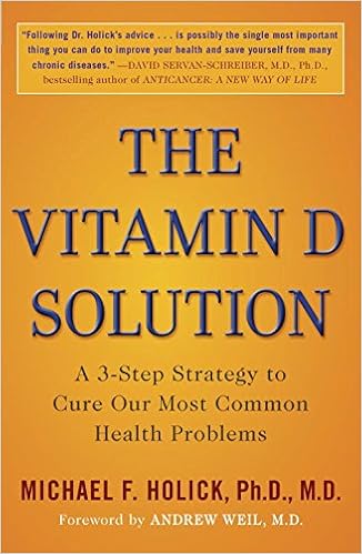
 The Vitamin D Solution

http://www.bumc.bu.edu/endo/faculty/holick/

Saturday, January 18, 2014

A Critical Review of the Classic Metaphyseal Lesion: Traumatic or Metabolic?

http://www.ajronline.org/doi/abs/10.2214/AJR.13.10540

David M. Ayoub, Charles Hyman, Marta Cohen and Marvin Miller

ABSTRACT :

OBJECTIVE. The purpose of this study was to review the hypothesis that classic metaphyseal lesions represent traumatic changes in abused infants and compare these lesions with healing rickets.

MATERIALS AND METHODS. Using a PubMed search, a multidisciplinary team reviewed studies that reported the histopathologic correlation of classic metaphyseal lesions. Selective studies of growth plate injury and rickets were cross-referenced.

RESULTS. Nine identified classic metaphyseal lesion studies were performed by the same principal investigator. Control subjects were inadequate. Details of abuse determination and metabolic bone disease exclusion were lacking. The presence of only a single radiology reviewer prevented establishment of interobserver variability. Microscopy was performed by two researchers who were not pathologists. Classic metaphyseal lesions have not been experimentally reproduced and are unrecognized in the accidental trauma literature. The proposed primary spongiosa location is inconsistent with the variable radiographic appearances. Classic metaphyseal lesions were not differentiated from tissue processing artifacts. Bleeding and callus were uncommon in spite of the vascular nature of the metaphysis. The conclusion that excessive hypertrophic chondrocytes secondary to vascular disruption were indicative of fracture healing contradicts the paucity of bleeding, callus, and periosteal reaction. Several similarities exist between classic metaphyseal lesions and healing rickets, including excessive hypertrophic chondrocytes. “Bucket-handle” and “corner fracture” classic metaphyseal lesions resemble healing rickets within the growth plate and the perichondrial ring, respectively. The age of presentation was more typical of bone fragility disorders, including rickets, than reported in prior child abuse series.

CONCLUSION. The hypothesis that classic metaphyseal lesions are secondary to child abuse is poorly supported. Their histologic and radiographic features are similar to healing infantile rickets. Until classic metaphyseal lesions are experimentally replicated and independently validated, their traumatic origin remains unsubstantiated.

Thursday, September 20, 2012

Adoption halted as court told baby milk led to 'innocent' couple being accuse of abuse


Vitamin supplements in baby milk may have led an innocent couple being condemned for battering their newborn son, a top family judge has heard

Adoption halted as court told baby milk led to 'innocent' couple being accuse of abuse
Adoption halted as court told baby milk led to 'innocent' couple being accuse of abuse Photo: ALAMY
The boy, who cannot be named, was taken away from his parents and was poised to be adopted after multiple broken bones were put down to child abuse.
But Lord Justice McFarlane halted the process yesterday after hearing how an extraordinary combination of medical events could have led to a case of congenital rickets being overlooked.
The parents, who have fought a three-year custody battle, have been given a final chance to get their son back.
It came after lawyers had what they described as a "light bulb moment" and understood the full significance of the child's medical records.
Michael Shrimpton, for the family, who are from the north of England, told the Court of Appeal in London that there is evidence that the boy was born with a Vitamin D deficiency, inherited from his mother, leading to "soft bones" and rickets.
It suggests that the broken bones could have occurred during his difficult forceps birth, or even in the womb.
Blood tests to check for signs of vitamin deficiency, when the boy was four weeks old were normal.
But the court heard hat it is possible that it was "masked" by the formula milk given to him by his mother – which contained Vitamin D supplements.
He added that there was "striking" evidence of severe abnormalities in the functioning of the baby boy's liver, an organ instrumental in processing Vitamin D.
The judge temporarily halted the adoption process and ordered urgent medical reports.
Having a child taken away is an “exceptionally awful” ordeal, he remarked, adding that it was essential to examine whether the Vitamin D deficiency explanation for the boy's injuries was "more than an intellectual possibility".
He also noted that there was no evidence of emotional difficulties, domestic violence, alcohol or drug abuse, or any signs of dysfunction within the family, to indicate a risk of child abuse.
Mr Shrimpton said that one of the country's top endocrinologists, Professor Stephen Nussey, who has carried out pioneering work on the causes and effects of Vitamin D deficiency, will be instructed to carry out that task if he is available at short notice.
Observing that medical knowledge on the causes of infant injuries is in a state of constant movement, the barrister added: "This is an important case. It is starting to take on the appearance of a leading test case".
After hearing expert evidence in June last year, a judge at Sheffield High Court ruled that one or other of the parents must have been responsible for the baby's injuries. The same judge refused to change her mind earlier this year and freed the boy for adoption.
However, Lord Justice McFarlane observed: "Medical knowledge of how some children may have bones that are more susceptible to injury than normal children has moved on".
Emphasising the extreme urgency of the case in light of plans for the boy's imminent adoption, the judge gave the parents 28 days to obtain a report from Professor Nussey, or another expert, in support of their case.
The local authority involved in the case had informed the Appeal Court that suitable adoptive parents have already been found for the boy but no further steps in the process would be taken prior to the court ruling on the case.
The case will return to the Appeal Court once the expert medical report has been obtained.

Monday, April 9, 2012

Murder-suicide over false child abuse claims

The family of a couple whose lives tragically ended in a murder-suicide after they were mistakenly accused of child abuse is finally sharing their story.
Almost four years after finding their daughter and son-in-law dead in their own home, Jackie and Paul Cuin spoke to the Denver Post, which obtained medical, social services and police investigations records relevant to their case.
In 2008, Alyssa O'Shell was taken from her parents, Dave and Tiffany, when she was just three months old. Doctors had found 11 broken bones in the infant's legs, prompting suspicions of abuse. Shocked, Dave and Tiffany were helpless as child protective services gave their only baby to a foster mother.
Insisting they were innocent, Alyssa's parents countered the allegations, researching the Internet for other explanations. Maybe she had brittle bone disease, they suggested. But doctors said the little girl's broken bones were "highly suggestive of nonaccidential trauma," according to a hospital report, and would have come from violently twisting her legs.
In the two weeks that followed, Dave and Tiffany, both police officers in Henderson, Colo., met with lawyers and searched for ways to prove their innocence and bring Alyssa back to their family.
Dave, after admitting he'd once playfully picked Alyssa up by her legs to kiss her stomach, was especially scrutinized, and began to think he would be arrested for felony child abuse. As each day passed, he grew more despondent. Tiffany had begun to wonder if divorcing her husband was the only way to keep her baby.
Tiffany's parents, Jackie and Paul Cuin, were also being investigated, as they often watched Alyssa while Dave and Tiffany were at work.
On June 30, 2008, Tiffany and Dave were each to meet with their respective lawyers and a criminal investigator. The last appointment was at 1 p.m. Jackie grew worried when she never heard from her daughter, who had agreed to call throughout the day with updates. At 3 p.m., she called Jackie. No answer.
At 5 p.m., she drove to the couple's home, where she saw both cars in the garage and the dog on the porch. Scared to go inside the house, she instead drove home and returned with her husband, Paul, who agreed to go inside.
"I had already dialed 911 when I heard him scream," Tiffany recalled to the Denver Post.
Inside, Paul found both Tiffany and Dave dead in their bedroom. They hadn't been at the appointments.
The night before, Dave reportedly shot his wife while she slept, then took his own life, sticking two handguns in his mouth and pulling both triggers.
Alyssa's parents were dead, but meanwhile, doctors were running more tests on the little girl. Her foster mother had expressed concerns about her lack of development, just like Tiffany and Dave had once done.
A doctor noted that Alyssa "makes sounds, smiles and laughs," but "does not grasp objects well," and had trouble moving her limbs and controlling her head. She also tended to keep her thumbs in her palms.
All the signs pointed to a genetic illness.
On July 9, Alyssa was diagnosed with spinal muscular atrophy, which weakens limbs and is associated with broken bones. The Cuins reached out to a specialist at a national spinal muscular atrophy association to confirm that Alyssa's broken bones could be attributed to the disease.
"I have spent three hours reviewing the xrays and the reports," said Dr. Gary Chan in an email to Jackie. "The fractures are consistent with subjects with SMA.
"I would guess some of these fractures may occur at time of delivery, but most occurred after birth from normal handling. It does not surprise me that Alyssa had fractures noted at 3 months of age."
In infants, spinal muscular atrophy is usually fatal, as it was for Alyssa. She died on October 28, 2008, just seven months old.
The Cuins, who were cleared of all abuse allegations and granted custody of Alyssa for the rest of her short life, sued the child protection team but lost.
Still angry, Paul says the social services departments become "overzealous," and should focus on reuniting families when it is appropriate.
"They didn't in this case," he told the Denver Post. "They literally tore the family apart. And you have no recourse. You have to prove you're innocent to get your child back."
Child and family services director Darwin Cox told the newspaper the hospital where Alyssa was referred from is renowned for its child-abuse expertise.
"This was a terrible, terrible tragedy," Cox said. "We did the job we could with the best information we had at the time."
rmurray@nydailynews.com


Friday, April 6, 2012

Doctors Say Rickets Is Back

By Melissa Schorr
B O S T O N, Aug. 10

An exclusive diet of breast milk could be putting some babies at risk for vitamin D deficiency and thus at risk for rickets, a bone-weakening disorder that has been virtually nonexistent in the United States for decades.

Doctors nationwide are reporting a small spike in the number of breast-fed babies developing nutritional rickets, Rickets is caused by a lack of vitamin D, which is crucial for strong bone development. It can result in stunted growth, broken bones and bow legs in children.

Breast milk, though an excellent source of nutrition overall, provides little vitamin D. Babies can get the vitamin instead from a splash of sunlight or from a daily supplement.

But rickets may be on the rise, researchers believe, because of the renewed popularity of breast-feeding, along with doctors’ failure to properly prescribe vitamin D supplements to breast-feeding moms.

Dark Skin, High Risk At special risk are dark-skinned babies, whose pigmentation prevents them from using sunlight to produce the necessary vitamin D.

But babies of any race who aren’t getting enough vitamin D from their diet and aren’t getting a daily dose of sunlight are potentially at risk of developing rickets, researchers say.

Doctors at two medical centers in North Carolina report in this month’s issue of the Journal of Pediatrics that they saw 30 cases of rickets in breast-fed black infants from 1990 though 1999, with more than half of the cases occurring in the last 18 months alone.

This June, researchers at the University of Texas Southwestern Medical Center in Dallas published a report in the journal Texas Medicine about a recent outbreak of rickets among dark-skinned infants who were breast fed, despite the presence of abundant sunlight in the state.

Statewide Surge Cases of rickets have been popping up around the country, from New York to Florida, notes Dr. Robert P. Schwartz, an endocrinologist at Wake Forest University School of Medicine in Winston-Salem, N.C., lead author of the study.

Schwartz decided to study the issue along with his colleagues, Dr. Shelley Kreiter, a pediatrician at Wake Forest, and Dr. Henry Kirkman of the University of North Carolina School of Medicine in Chapel Hill.

Because of growing concerns, the Centers for Disease Control will be putting out a bulletin on rickets this fall, a spokesman says.

“This is a disease that was here 100 years ago,” Schwartz says. “It’s back.”

History of Rickets

As late as the 1940s, rickets was a common childhood ailment, killing thousands of children annually until doctors learned that that vitamin D in milk and sunlight could largely prevent it.

Today, fortified milk and prepared baby formula both contain vitamin D, largely eliminating the problem.

But because breast milk contains much lower levels of vitamin D, babies exclusively fed on it may be at risk, especially if they have darker skin that blocks out sunlight; live in northern climates and receive less light or have moms with vitamin D deficiencies themselves.

The number of African-American women who are breast-feeding has been on the rise in the past decade, from about 5 to 22 percent, the Pediatrics study reports.

“We’re glad to see that more minority women are breast-feeding, and we don’t want them scared away because they hear their breast milk is not sufficient,” says Carol Huotari, manager for the Center for Breastfeeding Information at La Leche League International, a Schaumburg, Ill.-based group that promotes breast-feeding. “It’s a simple thing to provide vitamin D supplements.”

“We don’t want to come across as against breast-feeding,” Schwartz concurs. “But we think all infants should be supplemented with vitamins.”

Since January, Schwartz’ group has provided supplements to more than 700 women in North Carolina who are exclusively breast-feeding. The supplements are paid for by the federal Health Resources and Services Administration’s Maternal and Child Health Block Grant program.

“Proving supplements to every baby seems like overkill,” says Huotari, “when it’s just a selected group of babies that would benefit.”

But Schwartz says many infants are potentially at risk and the cost has been minimal so far: about $1.50 per infant per month — far less than hospitalizing infants for fractures and broken bones.

Doctors Fail to Warn Besides the increase in breast-feeding contributing to this rise in rickets, pediatricians also may not be telling mothers to supplement the breast milk with vitamin D.

The Journal of Pediatrics study surveyed 400 North Carolina pediatricians and found that 16 percent weren’t prescribing vitamin supplements at all. Younger doctors, who may have never seen a case of rickets, were less likely to prescribe vitamin D than older ones.

“Doctors have not been led to believe it is essential,” Schwartz explains.

Schwartz says the American Academy of Pediatricians hasn’t established clear enough guidelines on supplementation.

In 1998, the academy recommended vitamin D supplements be used for breast-fed infants with dark skin or inadequate exposure to sunlight, but other earlier policy statements still in effect haven’t provided a clear mandate for vitamin use.

“The nice thing is, this disease doesn’t have to occur at all,” Schwartz says. “This is 100 percent preventable.”

http://abcnews.go.com/Health/story?id=118054&page=1#.T36eC6tSSj_

Thursday, April 5, 2012

What is Hypophosphatasia (HPP)?


Hypophosphatasia is an inherited metabolic (chemical) bone disease that results from low levels of an enzyme called alkaline phosphatase (ALP). Enzymes are proteins that act in the body's chemical reactions by breaking down other chemicals. ALP is normally present in large amounts in bone and liver. In hypophosphatasia, abnormalities in the gene that makes ALP lead to production of inactive ALP. Subsequently, several chemicals - including phosphoethanolamine, pyridoxal 5'-phosphate (a form of vitamin B6) and inorganic pyrophosphate - accumulate in the body and are found in large amounts in the blood and urine of people with Hypophosphatasia. It appears that the accumulation of inorganic pyrophosphate is the cause of the characteristic defective calcification of bones in infants and children (rickets) and in adults (osteomalacia).
Nevertheless, the severity of hypophosphatasia is remarkably variable from patient-to-patient. The most severely affected fail to form a skeleton in the womb and are stillborn. The most mildly affected patients may show only low levels of ALP in the blood, yet never suffer bony problems.

In general, patients are categorized as having "perinatal", "childhood" or "adult" hypophosphatasia depending on the severity of the disease, which in turn is reflected by the age at which bony manifestations are first detected. Odontohypophosphatasia refers to children and adults who have only dental, but not skeletal, problems (premature loss of teeth).

The x-ray changes are quite distinct to the trained eye. Similarly, the diagnosis of hypophosphatasia is largely substantiated by measuring ALP in the blood (a routine test) that is low in hypophosphatasia. However, it is important that the doctors use appropriate age ranges for normals when interpreting an ALP level.

Prevalence

It has been estimated that severe forms of hypophosphatasia occur in approximately one per 100,000 live births. The more mild childhood and adult forms are probably somewhat more common. About one out of every 200 individuals in the United States may be a carrier for hypophosphatasia

Prognosis

The outcome following a diagnosis of hypophosphatasia is very variable. In general, the earlier the diagnosis is made the more severe the skeletal manifestations. Cases with severe, not mild, deformity at birth almost always have a lethal outcome within days or weeks. When the diagnosis is made before six months of age, some infants have a downhill and fatal course, others survive and may even do well. When diagnosed during childhood, there can by presence or absence of skeletal deformity from underlying rickets, but premature loss of teeth (less than five years of age) is the most common manifestation. Adults may be troubled by recurrent fractures in their feet and painful, partial fractures in their thigh bones.

Symptoms

Depending on the severity of the skeletal disease, there may be deformity of the limbs and chest. Pneumonia can result if chest distortion is severe. Recurrent fractures can occur. Teeth may be lost prematurely, have wide pulp (inside) chambers, and thereby be predisposed to cavities.

Inheritance Factors

The severe perinatal and infantile forms of hypophosphatasia are inherited as autosomal recessive conditions. The patient receives one defective gene from each parent. Some more mild (childhood or adult) hypophosphatasia cases are also inherited this way. Other mild adult and odonto hypophosphatasia cases seem to be inherited in an autosomal dominant pattern (the patient gets just one defective gene, not two, transmitted from one of his/her parents). In this form, mild hypophosphatasia can occur from generation-to-generation. The perinatal form of hypophosphatasia can often be detected during pregnancy by ultrasound and by measuring ALP activity in chorionic villus samples from amniocentesis.

Individuals with hypophosphatasia and parents of children with hypophosphatasia are encouraged to seek genetic counseling to explain the likelihood and severity of hypophosphatasia recurring in their families.

Treatments

As yet, there is no cure for hypophosphatasia and no proven medical therapy. Some medications are being evaluated. Treatment is generally directed towards preventing or correcting the symptoms or complications.

Expert dental care and physical therapy are recommended. An orthopaedic procedure called "rodding" may be especially helpful for adults with painful partial fractures in their thigh bones. Severely affected infants may manifest increased levels of calcium in their blood that may be treated with calcitonin and certain diuretics. Doctors should avoid the temptation to give calcium supplements or vitamin D unless there is clear-cut deficiency.

Contributing Medical Specialist
Michael P. Whyte. M.D.
Medical Director
Center for Metabolic Bone Disease and Molecular Research
Shriners Hospital
St. Louis, Missouri

Sunday, March 11, 2012

COMMON PEDIATRIC BONE DISEASES-APPROACH TO PATHOLOGICAL FRACTURES

COMMON PEDIATRIC BONE DISEASES-APPROACH TO PATHOLOGICAL FRACTURES

General Presentation

Background: It is not uncommon for children to present with fractures after experiencing trauma. However, children may also present with pathological fractures, which are fractures that occur in abnormal bones and typically occur during routine activity or after minor trauma. It is important to be able to distinguish between traumatic fractures and pathological fractures as the prognosis and treatment can vary quite considerably. One also MUST consider non-accidental injury in the child that presents with multiple unexplained fractures. Non-accidental injuries are discussed in a separate article on this site.
These fractures will generally present as localized pain and tenderness over the involved bone. Refusal to weight bear in the younger, non-verbal child is also very common.
Pathophysiology: Although there are many potential etiologies for weakened bone manifesting as pathological fractures, it is simple to divide it into three categories with common etiologies:
1)    Metabolic bone disease- eg. Rickets
2)    Bone tumours – eg. Benign tumours (non-ossifying fibroma and osteochondroma) and malignant tumours (osteosarcoma and Ewing’s sarcoma)
3)    Connective tissue bone disease- eg. Osteogenesis imperfecta
Metabolic Bone Disease: Normal bone growth and mineralization requires adequate calcium and phosphate. There are numerous etiologies of rickets, including nutritional deficiencies (calcium, phosphate and vitamin D), drug induced, renal pathology, and tumours. The end result and clinical manifestations of rickets are secondary to the failure of calcification of the growth plate cartilage because of a deficiency of either calcium or phosphate. Rickets is defined as deficient mineralization at the bone’s growth plate whereas osteomalacia refers to impaired mineralization of the bone matrix. Rickets is relatively common in children, especially those who have poor dietary intake, poor absorption, increased excretion of calcium, phosphate, or vitamin D, premature infants or breast-fed infants who are not supplemented with vitamin D. Regardless of whether it is calcium or phosphate deficient rickets, the typical clinical findings associated with Rickets include:
  • Skeletal Findings(see Figure 1):
    • Delayed closure of the fontanelle
    • Parietal and frontal bossing
    • Craniotabes (soft skull bones)
    • Rachitic rosary (enlargement of the costochondral junction such that there is beading across the anterolateral aspects of the chest)
    • Widening of the wrists; bowing the distal radius and ulna
    • Progressive bowing of the femur and tibia
  • Extraskeletal Findings(vary depending on the primary mineral deficiency):
    • Calcipenic rickets – hypoplasia of dental enamel, decreased muscle tone, delayed achievement of motor milestones, hypocalcemic seizures, increased sweating
    • Phosphopenic rickets – dental abscesses
Bone Tumors: Tumours, regardless of whether they are benign or malignant, can cause pathological fractures by growing and replacing the normal tissue of bone. This results in an abnormal, weakened bone more prone to fractures. In children, benign bone tumours are fairly common, but are often asymptomatic and discovered incidentally during evaluation for trauma or another condition, and thus, the true incidence is unknown. If they are symptomatic, they may present with localized pain, swelling, deformity or a pathological fracture. Most benign tumours generally present during the second decade. Two examples of common benign tumours which can present as pathological fractures include non-ossifying fibroma, and osteoid osteomas.
Malignant bone tumours account for 5% of all pediatric malignancies, with the peak occurrence between the ages of 10-24. The two most common malignant bone tumours in children are Ewing’s sarcoma and osteosarcoma which collectively make up 90% of the pediatric bone tumours. Ewing’s sarcoma is more common within the first 10 years, and then osteosarcoma becomes more common. The cancers often arise in the pelvis, femur, tibia and humerus. These can commonly present with symptoms of pain and swelling, which may be worse with exercise or at night, and sometimes the first signs may be due to a pathological fracture.
Connective Tissue Disease: Osteogenesis imperfecta (OI) is an inherited connective tissue disorder commonly known as “brittle bone disease” which can manifest in a wide spectrum, from mild to lethal forms. It is usually due to a deficiency of normal Type I collagen, which is an organic component necessary for proper bone formation.  Although its incidence is estimated to approximately 0.005%, it is important to include this disease in the differential diagnosis because it can present early in children as numerous and recurrent pathological fractures. The common clinical manifestations of OI include:
  • Multiple and/or atypical fractures
  • Short stature
  • Scoliosis
  • Basilar skull deformities
  • Wormian bones (irregular, small bones along the cranial sutures)
  • Blue sclera
  • Hearing loss
  • Opalescent teeth that wear quickly
  • Increased laxity of ligaments and skin
  • Easy bruising
  • Accelerated osteoporosis

Questions to Ask

  • How did the fracture occur – How, when, where? – to determine if it was pathological or traumatic fracture
  • Has the child had any previous fractures or any other concurrent fractures? If so, can you describe them? – to help determine pathological versus traumatic fracture
  • What is the child’s diet like? To determine if nutritional deficiency Rickets may be the cause
  • Did the child have any previous bone pain?  If so, can you describe what makes it better or worse? Also, does it get worse at night? To check for potential bone tumors
  • Does the child have a fever? To rule out any potential infectious causes or malignancies (constitutional symptoms – fevers, weight loss, drenching night sweats)
  • Does the child have any other medical history? To rule out a secondary cause of the bone disease
  • Do you suspect that the child may have been abused? Who is the primary caretaker of the child?
  • Is the child taking any other medications?

Differential Diagnosis for Pathological Fracture

  • Rickets from Vitamin D deficiency
  • Osteogenesis Imperfecta
  • Renal Osteodystrophy
  • Osteomyelitis
  • Child abuse
  • Preterm birth resulting in osteopenia – neonates
  • Fibrous dysplasia
  • Osteomalacia
  • Copper deficiency – infants: first 6 months
  • Bone tumours and cancers
  • Chronic Vitamin A toxicity
  • Metabolic diseases – leading to calcium wasting and demineralization
  • Prolonged administration of prostaglandins, glucocorticoids, or methotrexate
  • Congenital syphilitic periostitis
  • Hypophospatasia
  • Juvenile Osteoporosis

Investigations and Management

1)    Rickets:
Laboratory findings:
  1. Elevated alkaline phosphatase – indication of impaired bone mineralizeation
  2. Serum phosphorous concentrations – usually low in hypocalcemic and hypophosphatemic rickets
  3. Serum calcium concentration – decreased only in hypocalcemic rickets
  4. Parathyroid hormone – usually elevated in hypocalcemic rickets, but usually normal in hypophosphatemic rickets
  5. 25-OH Vitamin D – low in vitamin D deficiency
  6. GFR and Creatinine – to determine kidney function
Radiographic Findings:
  1. Osteopenia
  2. Metaphyseal cupping  and fraying (See Figure 2)
  3. Physeal widening
  4. Enlargement of costochondral junction
  5. Bowing of long bones (See Figure 3)
Management:
  1. Oral doses of 5,000-15,000 IU/day of Vitamin for 4 weeks for Vitamin deficient Rickets
  2. Optimize calcium intake for hypocalcemic rickets
  3. Treat underlying primary cause of Rickets
2)    Bone Tumors (benign and malignant)
Laboratory Findings:
  1. If suspect malignancy: blood work including liver enzymes, CT chest, bone scan, bone biopsy, MRI of affected bone
Radiographic Findings:
  1. Benign: single lesion generally, sharp area of delineation, overlying cortex intact, sclerotic margins, no or simple periosteal reaction (See Figure 4)
  2. Malignant: multiple lesions often, poor delineation of lesion, loss of overlying cortex, extensive periosteal reaction, potential soft tissue involvement (See Figure 5)
Management:
  1. Benign: follow up with radiographs 4-6 months later
  2. Malignant: complete resection, chemotherapy, radiation
3)    Osteogenesis Imperfecta
Laboratory Findings:
  1. Elevated levels of serum alkaline phosphatase
  2. Hypercalciuria – magnitude reflects severity of disease
  3. C-terminal peptide (marker of bone formation) and C-telopeptide (marker of bone resorption) –can be higher
Radiographic Findings:
  1. Mild OI: Thin cortex and relatively few fractures with normal skull development
  2. More severe OI: hyperplastic callus formation (from thickened periosteum), shortened long bones with multiple fractures (See see Figure 6)
Management:
  1. Bisphosphonates

Conclusion

Children who present with pathological fractures always require a thorough evaluation. It is important to keep an open mind as to the various causes of pathologic fractures and to always rule out non-accidental injury.

References

1)        Beary J, Chines A. Clinical features and diagnosis of osteogenesis imperfecta. (Last Updated June 15, 2010) In: UpToDate, Tepas E (Ed), UpToDate, Wellesley, MA, 2010.
2)        Scheri S. Differential diagnosis of the orthopedic manifestations of child abuse. (Last Updated Dec. 3, 2008) In: UpToDate, Wiley E (Ed), UpToDate, Wellesley, MA, 2010.
3)        Rauch F. Overview of Rickets in Children. (Last Updated August 11, 2010). In: UpToDate, Hoppin A (Ed), UpToDate, Wellesley, MA, 2010.
4)        Tis J. Overview of benign bone tumors in children and adolescents. (Last Updated September 28, 2010) In: UpToDate, Torchia, M (Ed), UpToDate, Wellesley, MA, 2010.
5)        Kliegman R, Behrman, Jenson H, Stanton B. Nelson Textbook of Pediatrics, 18th ed. Philadelphia: Saunders, 2007.
6)        Benson M, Fixsen J, Macnicol M. Children’s Orthopaedics and Fractures, 3rd ed. New York: Springer, 2010.
7)        Kirpalani A, Babyn P. Imaging in Osteogenesis Imperfecta. eMedicine (Last Updated August 5, 2008). Available from http://emedicine.medscape.com/article/411919-print [Accessed on March 5, 2011]
8)        Rijn R, McHugh K. Rickets Imaging. eMedicine (Last Updated March 18, 2009). Available fromhttp://emedicine.medscape.com/article/412862-print [Accessed on March 5, 2011]
9)        Dugani S, and Lam D. Toronto Notes. Toronto Notes Medical Publishing Inc. 2009
10)     Jenny C. Evaluating Infants and Young Children with Multiple Fractures. Pediatrics. 2006; 118(3):1299-303.
11)      Adam A, Dixon A. Adam: Grainger & Allison’s Diagnostic Radiology, 5th ed. Philadelphia: Churchill Livingstone, An Imprint of Elsevier, 2008.

Acknowledgements

Written by: Teresa Liang
Edited by: Anne Marie Jekyll, MD (Pediatric Resident)

Images

(Image from Rijn R, McHugh K. Rickets Imaging. eMedicine (Last Updated March 18, 2009). Available fromhttp://emedicine.medscape.com/article/412862-print [Accessed on March 5, 2011])

(Image from Rijn R, McHugh K. Rickets Imaging. eMedicine (Last Updated March 18, 2009). Available fromhttp://emedicine.medscape.com/article/412862-print [Accessed on March 5, 2011])
(Adapted from Adam A, Dixon A. Adam: Grainger & Allison’s Diagnostic Radiology, 5th ed. Philadelphia: Churchill Livingstone, An Imprint of Elsevier, 2008)
(Adapted from Adam A, Dixon A. Adam: Grainger & Allison’s Diagnostic Radiology, 5th ed. Philadelphia: Churchill Livingstone, An Imprint of Elsevier, 2008)
(Image from Kirpalani A, Babyn P. Imaging in Osteogenesis Imperfecta. eMedicine (Last Updated August 5,2009). Available from http://emedicine.medscape.com/article/411919-print [Accessed on March 5, 2011])